Fertilization and Meiosis, (interacting opposites) [pdf]
Eggs are encased
 |
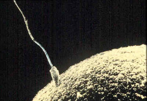
Fig. 1: Sperm contact.
Chromosome union ~10 hr later.
sperm head ~5x3 μm, tail 50 μm
human egg 100 μm diameter
(human hair ~90 μm diameter) |
in a protective lining called the zona pellucida to help them traverse the fallopian tube without sticking,
but they also emit a chemical to help sperm find them and have attachment sites for sperm to latch onto. There are usually several or many sperm contacting an egg at the same time, so the egg has to have fast lock-out mechanisms for preventing penetration from all others after the first. It takes three days to travel down the fallopian tube, and in that time there are typically six cell divisions producing an embryo with 26 = 64 cells. But it is in the uterus after the egg breaks out of the zona pellucida that the embryo receives its first nutrition and can grow in size. If all goes well, which it often doesn't, implantation occurs on day five or six.
Definitions:
oocyte: mature egg
zygote: fertilized egg
blastocyst: shell embryo
at about 4 days
ectopic: tubal (pregnancy)
gamete: egg or sperm
somatic: non-sex cells
ploid: number of sets
of chromosomes
diploid: two sets
haploid = monnoploid, one
stem cell: divides indefinitely
mitosis: normal cell division
meiosis: gamete producing
spindles: attachments for
microtubules to pull
chromosomes apart
polar body: discarded DNA
during egg meiosis
zona pellucida: wrapping
around oocyte and
early embryo
chromatid: duplicate DNA
chromatin: double-coiled DNA
amino acid: building blocks
for making proteins,
specified by a 4 base
sequence along DNA
transcription: using the DNA
base-sequence code
mRNA: messenger RNA
transcribed from DNA
translation: mRNA read to
make protein in cell
metaphase: cell diving
interphase: cell not dividing
IVF: petri dish fertilization
implantation: natural embryo
embeds in uterine
lining, or IVF embryo
placed in uterus
fallopian tubes: 7-12 cm long
1 nm = 1 millionth of a mm
1 μm = 1 thousandth mm
|
|
 |
Fig. 2: DNA helix.
2 m x 2 nm if straight
¼ trillion base pairs
3 bases per code
avg 485 codes/gene
≈30,000 genes for proteins |
That's the big picture, but the subject of this page is what happens to the DNA. Every cell, except for the outer skin, hair, finger nails and red blood cells, contains a complete identical set of DNA with a quarter trillion base pairs. And that has to be copied exactly before any cell division can occur. The usual process for replacing worn out cells or for growth of embryos is called mitosis as depicted in Figure 4. This happens six times while the embryo passes down the fallopian tube and eventualy some 40 trillion times by adulthood.
But the process called meiosis for making eggs and sperm is different. It involves one duplication of the DNA followed by two cell divisions as depicted in Figures 5 and 6. And what's particularly complicated and interesting is when and how the DNA of egg and sperm combine to restore the DNA to a complete diploid set.
At the time of sperm contact pictured in Figure 1, the egg's macro structure is more like Figure 7. The small polar body is cast off DNA from the bottom part of Figure 5 for Meiosis I. Internally in Figure 1, the egg still contains a full diploid set of 2x23 chromosomes, not just one set of 23.
It is only after sperm contact that the egg begins to execute Meiosis II - with no DNA duplication this time, and using spindles from the sperm to anchor the microtubules for pulling the chromosomes apart. The chromosomes are split down to only one set of 23. And this second cell division produces a second polar body, like Figure 7, with more cast off DNA. Then, finally, the 23 father chromosomes can combine with a corresponding set of 23 mother chromosomes to form a normal diploid nucleus with 2x23 chromosomes. This union occurs about 10 hours after the initial sperm contact pictured in Figure 1.
Chromosomes and Chromatids:
Human DNA is arranged into two sets of 23 chromosomes, for a total of 46, as depicted in Figure 3. This is what can be discerned in microscopes just before cell division. Note that the drawing is flat two-dimensional showing chromosomes lying on the page with their x-shaped structure showing after DNA duplication. But cells are three dimensional so that when viewed in a microscope their chromosomes may overly each other or may be tilted sideways so that the "x" structure can not be seen, the smallest Y chromosome being particularly difficult to discern. It takes many observations to deduce the arrangement drawn in this figure, and the final conclusive count of number of chromosomes in humans including the sex determining X and Y wasn't until 1956.
 |
Fig. 3: Graphic depiction of the 2x23=46 human chromosomes ordered by size.
Blue indicates paternal, red maternal. A gene on Y sets the developmental path towards maleness.
Chromosome 1 contains approximately 1873 genes (on each chromatid) and Y approximately 122.
(In other primates human gene #2 occurs in two smaller pieces giving total count of 2x24=48 chromosomes.)
(Chromosomes in dog 2x39=78, cat 2x19=38, jack jumper ant 2x1=2 in female but 1 in male)
|
Though it's what is always seen in microscopes, the "x" structure of the chromosomes is not really normal. It is due to the duplication of DNA that occurs just before cell division. Each side of an "x" is identical and called a chromatid. There are 92 chromatids. The cross point of the "x" is called a centromere, and is the point where the two chromatids will be pulled apart when cell division occurs. This dividing phase of a cell's life cycle is called metaphase. Most of the time the cell is not dividing and is said to be in interphase.
The reason these dual-strand metaphase chromosomes are visible in a light microscope is that their DNA strands are unusually highly coiled to be short, compact and non-interfering with each other. This gives them a diameter of about 700 nm. That's bigger than the wavelength of visible light, a requirement for being able to deflect light waves and be seen.
Mitosis, Normal Cell Division:
Figure 4 depicts what happens next in normal mitotic cell division. Microtubules attach to the centromeres and pull the double-strand chromosomes apart. But as soon as the chromatids separate they are called chromosomes. Each daughter cell receives 23 pairs of single-strand chromosomes.
Then as the cell wall divides into two cells and nuclei congeal, the chromosomes disappear. This is because the unusually coiled state unwinds to just two levels of coiling in the normal configuration of DNA called chromatin. The diameter of chromatin is 30 nm, so even the shortest 450 nm wavelength of visible light just flows around this fiber with no effect.
Cells spend most of their life cycle in this interphase state with single-strand chromosomes invisible in light microscopes. The strands are also normally so twisted and entangled that even an electron microscope can not distinguish individual chromosomes. Cell nuclei are typically 6 μm in diameter but contain about 2 cm of the double coiled DNA called chromatin.
And to be active for making messenger RNA, individual gene sections of a chromosome unwind from the 6 μm diameter of chromatin to 50 times longer bare double helix of Figure 2. This then separates into its two backbone strands for code reading to make mRNA, which moves out of the nucleus into the cell for making the protein. During interphase the chromosomes, part wound as chromatin and part unwound to active DNA, are highly intertwined inside the nucleus.

Fig. 4: Mitosis
normal cell division
|

Fig. 5: Meiosis I
for egg 2 hr before ovulation
(one daughter cell very small and discarded)
(first state held since 7 month fetal stage)
|

Fig. 6: Meiosis II
for egg 2 hr after sperm contact
(one daughter cell very small and discarded)
(Sperm chromosomes not shown, but they combine with remaining egg chromosomes
about 10 hr after initial sperm-egg contact.) |
Meiosis, the Making of Egg and Sperm
Meiosis uses two cell divisions to achieve two things - first a randomization of genes on each chromosome (Fig. 5, meiosis I), and then halving of the number of chromosomes to one set of 23 (Fig. 6, meiosis II).
After meiosis the single copy of a chromosome is not an exact copy solely from either mother or father. Instead, the genes on a given chromosome may independently be from either parent. Take chromosome 1 with its 1873 genes, for instance. The first gene could be from the father, the second from the mother, the third from the father, etc. This is indicated in Figure 5 by the mixed red-blue coloring of the paired double-strand chromosomes. There are a lot of possibilities, 21873 = 7 followed by 823 zeros for chromosome #1 after gene mixing.
This genetic variety is perhaps the most fundamental part of sexual reproduction, apparently facilitating adaptability for survival of species. It's very basic and evolved a very long time ago. All sexually reproducing species of animals, plants and yeasts use the same basic enzymes for achieving gene randomization during meiosis.
In men meiosis is prolific. Five billion stem cells continually divide to produce 300 million sperm per day. This tremendous amount of DNA copying causes some vulnerability to error mutations.
In women, though, the stem cell stage stops while she is still a fetus at seven months with about seven million pre-egg cells, all suspended for 12-50 years at the beginning of meiosis I as in the top of Figure 5. Meiosis I egg cell division does not complete until just before ovulation, and the long wait causes vulnerability to microtubule breakage and thus genetically infertile eggs or birth defects.
Cytoplasmic Preparation
In addition to completing meiosis I, another thing that must proceed well in the last couple months and especially the last few hours before ovulation is preparation of special stores of cytoplasm. This is a period of very high metabolic activity, achieved partly with the aid of surrounding cells and, notably, even with the abnormally highly coiled, dual-strand chromosomes pictured in the upper half of Figure 5.
 |
Fig. 7: Mouse egg
with polar body
in zona pellucida |
The mature egg cell is enlarged by a factor of 500 in volume, diameter increasing from 10 to 80 μm.(ref) And when the meiotic cell divisions occur this store of egg cytoplasm is not divided in half. The cytoplasm is almost all retained by only one daughter cell. The other daughter cell is small, sloughed off and discarded. So the two cell divisions of meiosis produces only one large egg, called an oocyte, plus some small "polar bodies" that contain rejected chromosomes and very little cytoplasm. This oocyte has the energy supply and chemical factory components needed to divide into 64 smaller cells without the aid of external nutrition.
The sperm, on the other hand, is very small. It does not have to provide any energy supply or chemistry except just for its own survival for a couple days, ability to swim, and enzymes for dissolving through the zona pellucida. "Small" means that it has only about 6000 proteins.
Common Errors in Egg Development and Fertilization:
Diploid cells have two copies of every chromosome. There can be differences like for blue eyes versus brown eyes for a given gene, but the two copies of each chromosome are still basically the same. The pairs contain the same number, order and structure of genes. Cats and dogs have 2x19 and 2x38 chromosomes respectively, but are still diploid like humans.
Polyploidism occurs commonly in plants. Wild wheat is diploid, for instance, but commercially developed bread wheat is hexaploid with six copies of every chromosome.
Haploid cells have only one copy of every chromosome, are monoploid. These are the gamete cells, the egg or sperm.
Aneuploid means that the number of chromosomes is mixed up. In monomer embryos a chromosome occurs with only one copy instead of two. In trimer embryos there are three copies of a chromosome instead of two. Monomers tend to fail early after just a few cell divisions. Trimers have a high failure rate, too, but a few may survive. Down syndrome is trisomy 21, three copies of chromosome 21.
Aneuploidism can happen because of the long suspended state of pre-meiosis I in females. If a right-hand microtubule in Figure 5 becomes damaged, the chromosome it should be connected to will be drawn to the left instead of the right. And then depending on which side is the large egg or the small cast off polar body, the mature egg or oocyte will have either two copies or no copy of that gene. Fertilization then produces either three or one copy of the chromosome.
There is still another completely different form of oddity that can happen, though not very often. In Figure 5 or 6, meiotic cell division is supposed to retain almost all the cytoplasm for a large egg on one side and cast off almost no cytoplasm in a small polar body on the other side. In some one in a thousand cases, though, the polar body remains large, the macrostructure becoming a modified Figure 7 with two more or less equal, half-size eggs inside the zona pellucida.
Nothing unusual happens if only one of the half-size eggs becomes fertilized. Well, there may be reduced survival rate because of the loss of cytoplasm. But if both eggs in the zona pellucida become fertilized two things can happen. Most commonly, the two oocytes develop independently for three days, long enough break out of the zona pellucida in the uterus and become clearly independent. They are called identical half twins, a form of fraternal twins, but with more genetic similarity.
Very rarely, a pair of fertilized cells inside the zona pellucida will merge into a single embryo. There are a couple other ways this can happen, too, as from implanting too many IVF embryos, but it always leads to a difficult or very bad outcome. This is where impossible mixed blood types come from, or both sexes in one individual. There is even discussion in the literature about what fraction of each sex an individual will become based on the time delay between the two fertilizations. The freakiness is apparently stressful. A known chimer who wrote a life story book ended up committing suicide.
|